Cell Biology of RNA viruses
Head: Dr. Gabrielle Vieyres
Lipid droplets in viral infections
As obligate intracellular pathogens with a limited genome size, RNA viruses have mastered the skill of drawing resources from the host cell while outsmarting its defenses. Some of the strategies used are broadly conserved among pathogens. For instance, plus-strand RNA viruses reshuffle host cell membranes to form a replication organelle. This compartment concentrates viral and usurped cellular replication factors and isolates the viral replication machinery from the innate immune defenses. In addition to inducing new specialized compartments, viruses also exploit existing compartments of the host cell. This is the case of the endocytic compartment, associated with the entry of multiple viruses already over 40 years ago. More recently lipid droplets have emerged as a key organelle for the replication of a spectrum of pathogens, including viruses, bacteria and parasites. In many cases however, their exact role in the pathogen replication cycle and the machinery used by the pathogen to hijack this organelle are poorly understood. Lipid droplets are the main energy reservoir of the cell but also the source for structural and signaling lipids, all coveted resources for pathogens. Given this role as a metabolic hub, it is likely that their role in pathogen infection is still underestimated.
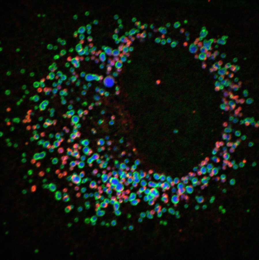
Within the InterACt network and based at the Heinrich Pette Institute, the junior research group “Cell Biology of RNA Viruses” examines the spatiotemporal dynamics of viral replication compartments, with a focus on RNA viruses and lipid droplets. Hepatitis C virus (HCV) is a particularly interesting model for this pathogen-host interaction: its morphogenesis relies on host lipid droplets, and its pathogenesis, in particular HCV-induced nonalcoholic fatty liver disease (NAFLD), involves the modification of this compartment. We have previously reported that hepatitis C virus hijacks a lipolytic pathway of the host hepatocyte to extract neutral lipids from the lipid droplets and support its own morphogenesis (Vieyres et al., PLoS Pathogens 2016; Vieyres et al., PLoS Pathogens 2020). We are further unraveling this interplay, at the level of individual lipid droplets, and monitoring the effect of HCV infection on this compartment. In this regard, we believe HCV research can also provide a precious insight into the functions and malfunctions of this organelle that is central in the current epidemics of fatty liver disease. Supported by the spectrum of competences from the InterACt Science Campus, and in synergy with the research group of Prof. Eva Herker at Marburg University,, we use our expertise in molecular virology and cell biology, combined with high-resolution fluorescence microscopy, live-cell imaging, unbiased image analysis methods and data mining, to highlight new interactions between RNA viruses and this organelle, and better understand their function in virus replication and pathogenesis. In the long term, targeting antiviral compounds to this central cell compartment or “closing the tap” that fuels viral replication might open new anti-infective therapeutic perspectives.
